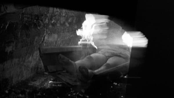
When I see the handle @mrs_angemi, (or @mrs anything)I immediately think workout selfies, baby pictures and flowers from doting hubby. I couldn’t be more wrong about Nicole Angemi‘s Instagram account, a collection of images from her work as an assistant pathologist. Despite the fact that her Instagram account has been shut down several times in the year since she started posting these images, and despite the dozens of times her images have been flagged and reported, she doesn’t post these photos for gratuitous purposes. Angemi’s photos are clearly meant to educate and enlighten people about a subject they are usually uncomfortable with – death and disease and its myriad causes and effects. Her aim is to share autopsies with the general public; she doesn’t believe that only those in the medical field should have access to these images and this knowledge. I couldn’t agree more – especially when you consider the number of inaccurate Hollywood autopsies there are on TV every week. I personally think she’s using social media in an ideal way, to share knowledge with people as opposed to sharing what your legs look like at the beach. If you’re into gross but fascinating stuff and interested in what strange things the human body can carry – like a massive ovarian cyst larger than an 8-month pregnant belly – then you should add yourself to the over 160K followers that @mrs_angemi currently boasts! Also, check out this awesome documentary feature on her via Motherboard…thank god they will never invent smell-o-vision!

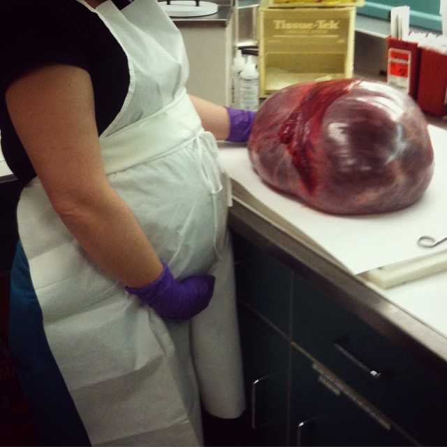 This is the ovarian cyst I was talking about…
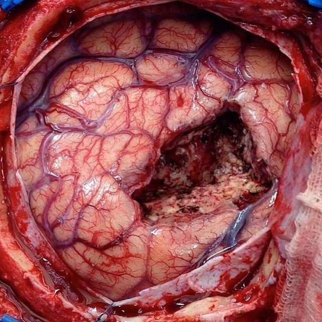
Open brain surgery!!! Here you can see the hole or defect where the surgeon removed a piece of brain. That is a huge chunk of brain right!!? You can actually live without pretty large chunks of brain depending what part of the brain was operated on. Brain surgery like this is commonly done to remove tumors and also parts of the temporal lobe for patients with severe epilepsy.
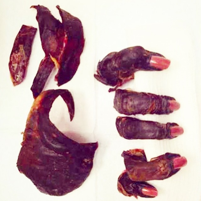
I always love these specimens. This lady’s hand was clearly dead. For a long time, to the point of mummification. Yet, she recently painted her nails just prior to having most of her hand surgically removed for gangrene. Got to maintain beauty at all times! 💅
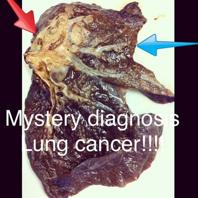
This is a total lung resection with a large cancerous tumor present in the bronchus or airway of the lung (red arrow). The tumor is so big it is causing an obstruction of the bronchus which led to a secondary infection causing the entire lobe of the lung to have pneumonia or lobar pneumonia (blue arrow). This is 100% attributed to smoking!!!!!!
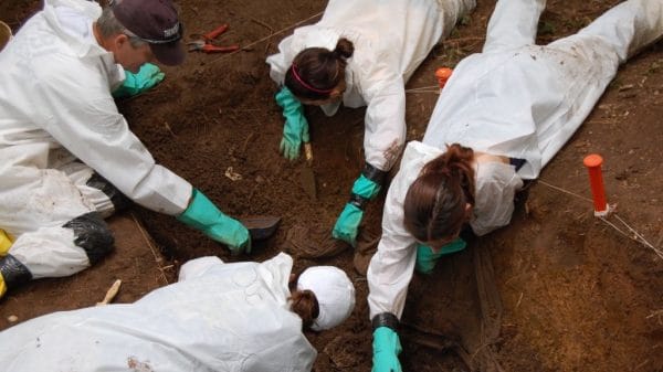
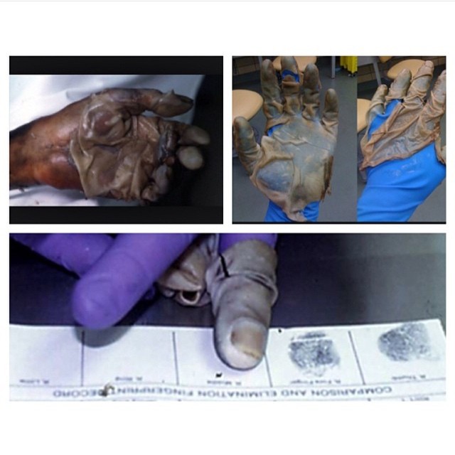 Forensic Friday!!!! This is another good Forensic Friday post from one of my old accounts. This was a very cool technique I learned at my time interning at the Medical Examiners Office that I thought was the coolest thing ever!!! When unknown dead bodies are received, we have to try to identify them so we can notify their next of kin. When a body has little to no clothing or identification on them, we fingerprint the body. When a body is dead for some time and begins to decompose, the skin undergoes changes in which the top layer of skin (epidermis) detaches from the bottom layer of skin (dermis). This is called skin slippage. If the unidentified body has begun decomposing, it is hard to take their fingerprints because their skin is literally slipping all over the place😁 The solution? Take off the dead guy’s slipped skin and put it on our hands as a glove so we can take the fingerprints!!!!! CREEPY!!!! But soooooo cool!!!! The top right photo shows the skin off the dead man from the top left photo on the gloves of the person performing the autopsy. The bottom photo shows this person taking fingerprints with the dead mans skin!!!!!
Fetal brain at 18 weeks.
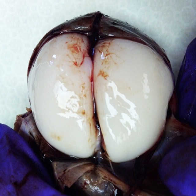
This is a brain!? Where are all the wrinkles??? The brain wrinkles are called the gyri which are the prominent portion of the folds and the sulci which are the grooves of the folds. These wrinkles or folds increase the surface area of the brain allowing for maximum brain power in our tiny skulls!!! These wrinkles start to appear later in fetal life, after 20 weeks and are gradually more prominent the older the fetus gets.